
Equine Thermal Imaging NOW AVAILABLE
Thermal Imaging has been honed and developed over the past thirty years, and scientifically proven in its benefits to vets and paraprofessionals. Veterinary Thermal Imaging provides a valuable addition to existing diagnostic tools such as X-rays, CT, MRI and Ultrasound scanning offered by your Vet. It also provides a visual management tool for owners and professionals monitoring or treating injury, illness or disease.
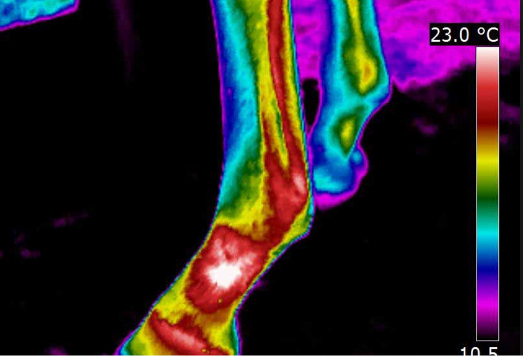
Thermal Imaging is very sensitive to changes in the muscular, vascular, skeletal and nervous systems, detecting temperature differences of less than 0.05oC which is 40 times more sensitive than the human hand.
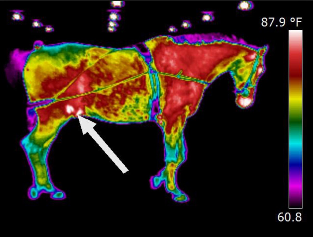
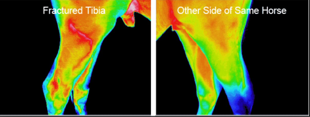
Thermal Imaging fills a gap in clinical diagnosis tools, and shows the animal's physiological state by graphically mapping skin surface temperature in response to changes in blood flow. In healthy animals, the thermal pattern on the skin is symmetrical. This is because skin blood flow is controlled by the sympathetic nervous system.
Clinical uses for Veterinary Thermal Imaging include:
Thermal Imaging is very sensitive to changes in the muscular, vascular, skeletal and nervous systems, detecting temperature differences of less than 0.05oC which is 40 times more sensitive than the human hand.
Thermal Imaging fills a gap in clinical diagnosis tools, and shows the animal's physiological state by graphically mapping skin surface temperature in response to changes in blood flow. In healthy animals, the thermal pattern on the skin is symmetrical. This is because skin blood flow is controlled by the sympathetic nervous system.
Clinical uses for Veterinary Thermal Imaging include:
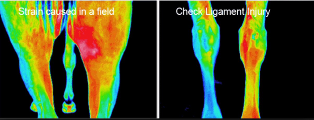
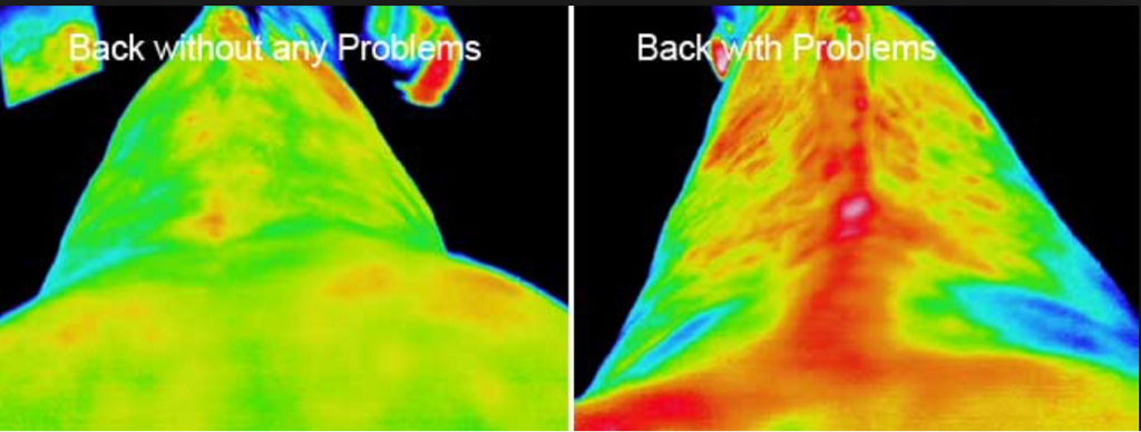
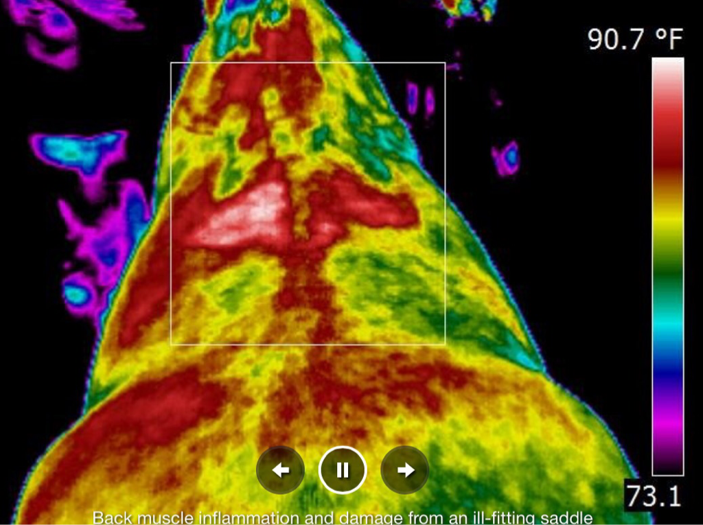
- Determing the extent of a diagnosed injury
- Detecting early lesions before they are clinically evident
- Identifying areas not previously identified where further diagnostic tests should be performed
- Monitoring the healing process before the animal is returned to work or training
|
|
|
|
|
|
